Canine Cardiopulmonary Filariasis.
Author: DR. GINO PICCININI veterinary dr.
Graduated from Bologna University-July 1978
Veterinary Surgeon in Nogara (VR) and Ostiglia (MN)
It is not necessary to the history of the heart filariasis, but as you can
see in table1-all. Para n. 31.d- (taken from an 18th century manuscript)
Heart worm was described: "... Lives in the heart of the dog, (true)
but rhe larvae, can be transported by the blood system to the subcutaneous
connective tissue, where they place themselves, giving them the nodule form
... " (incorrect).
The manual of pathological anatomy of domestic animals, by Prof. Kitt,
edited in the Autumn of 1895, states that "... the dog hosts many
parasites in its blood, of the Nematode type. The most important is the
filaria immitis or haematica, the heart worm, while the embryos, in a
numerous quantity, travel throughout the blood ..."
The researchers, Grassi and Noè, proved around the 1900 that the
larvae form of the filariae, passed from the mosquito to the dog during
a blood meal thanks to active mechanisms of swelling and folding of the
buccall wall (proboscides).
The Filaria Immitis LEIDY is threadlike, with a diameter of around 1 mm;
the female can reach up to 35 cm. long, while up to 16 cm in the male.
They are both of a whitish colour.
In the book of pathological and special treatments for domestic animals
(HUTYRA- MAREK), edited in 1929, the following was emphasised: Frequency,
Etiology, Development of the haematic filariae, Resistance of the micro-filariae,
Naturalcontamination, Pathogenesis, Pathological and anatomical lesions,
Symptoms and diagnosis.
|
|
|
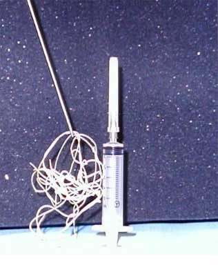
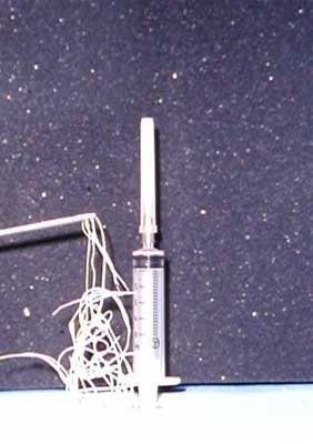
Macrofilariae (kept in 10% of formaldehyde) removed
from the right side Of the heart, during a post mortem on an 8 (eight)
year old, tawny coated male Boxer dog.
|
|