|
|
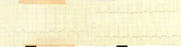
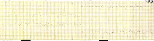
| Electrocardiogram. 2-year-old male Boxer dog. Infected with C.P.F. Initial right ventricular hypertrophy, deviation of the left axis, T wave-negative (5.1 mm), sinusal arrhythmy. I want to say (as follows) that all the significant elements to define E.C.G. are missing-for initial right ventricular hypertrophy. |
  |
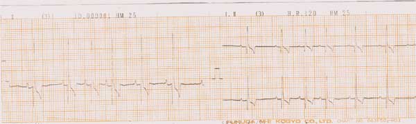
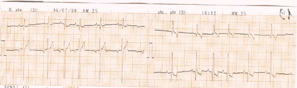
4-year-old female Boxer dog. C.P.F. First degree atrioventricular blockage. Left paw blocked and extension of the QT tract. This dog also had sub-aortic stenosis and ventricular hypokinaesthesia. |
up
|